|
| |
Ultra-400LAX
 Zenith
Microscopes
Zenith
Microscopes
Monocular/Binocular Biological Microscopes (Student Type)
Ultra-400LAX'
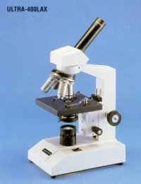
Zenith Ultra-400LAX Advanced Student Microscope
Ideal for ordinary and advanced level biology studies. The Zenith
Ultra-400 series are purpose built teaching instruments finished to a
very high standard, combining excellent optical performance and a very
reliable, modern, user-friendly design with a mechanical construction
designed to withstand the rigours or everyday heavy classroom use. All
models feature high quality DIN standard optics, anti-tamper safety
features included locked on eyepiece and stage clips, slip clutch on the
coarse focus movement to prevent over focussing and an adjustable focus
stop to prevent damage to glass microslides and objective lenses. Highly
recommended.
Standard Specification:
- Magnification Range x40, x100, x400,x1000
- DIN Achromatic Objectives x4 (0.10), x10 (0.25), x40R
(0.65),100R(1.25) oil immersion (R=retractable)
- x10 widefield eyepiece with pointer (18mm field)
- ABBE Condenser (N.A. 1.25) in spiral focussing mount with iris
diaphragm
- Quadruple Nosepiece Turret
- Built in 230v 20w illumination (colour corrected with daylight
blue filter)
| Accessories |
| WF-5 |
x5
DIN eyepiece |
| WF-16 |
x16
DIN widefield eyepiece |
| ME-10 |
x10
DIN widefield measuring eyepiece |
| CT-11 |
Mechanical
Stage |
| VA-2 |
Teaching
Head |
| VA-2TV |
CCTV
Adaptor (Mag. X2) |
| BS-3 |
Plano-concave
mirror in mount |
| SB-20 |
Replacement
240v 20w bulb |
|
Product Code: Ultra-400LAX
build_table('etistore',array('heultra400lax')); ?>
M
A microscope is an instrument for viewing objects that are too small to be seen by the naked or unaided eye. The science of investigating small objects using such an instrument is called microscopy. The term microscopic means minute or very small, not visible with the eye unless aided by a microscope. The microscopes used in schools and homes trace their history back almost 400 years.
The first useful microscope was developed in the Netherlands in the early 1600s.[1] Three different eyeglass makers have been given credit for the invention: Hans Lippershey (who also developed the first real telescope); Hans Janssen; and his son, Zacharias. The coining of the name "microscope" has been credited to Giovanni Faber, who gave that name to Galileo Galilei's compound microscope in 1625. (Galileo had called it the "occhiolino" or "little eye".)
The most common type of microscope—and the first to be invented—is the optical microscope. This is an optical instrument containing one or more lenses that produce an enlarged image of an object placed in the focal plane of the lens(es). There are, however, many other microscope designs.
1 Types
1.1 Optical microscopes
1.2 Electron microscopes
1.3 Scanning probe microscope
1.4 Other microscopes
2 External links
An optical light microscope
A stereo microscope is often used for lower-power magnification on large subjects."Microscopes" can largely be separated into three classes, optical theory microscopes, electron microscopes and scanning probe microscopes.
Optical theory microscopes are microscopes which function through the optical theory of lenses in order to magnify the image generated by the passage of a wave through the sample. The waves used are either electromagnetic in optical microscopes or electron beams in electron microscopes. The types are the Compound Light, Stereo, and the electron microscope.
Optical microscopes
Main article: Optical microscope
Optical microscopes, through their use of visible wavelengths of light, are the simplest and hence most widely used type of biology and geology.
Optical microscopes use refractive lenses, typically of glass and occasionally of plastic, to focus light into the eye or another light detector. Typical magnification of a light microscope is up to 1500x with a theoretical resolution of around 0.2 micrometres or 200 nanometers. Specialised techniques (e.g., scanning confocal microscopy) may exceed this magnification but the resolution is an insurmountable diffraction limit.
Various wavelengths of light are sometimes used for special purposes, for example, in the study of biological tissue.[2] Ultraviolet light is used to illuminate the object being viewed in order to excite a fluorescent dye which then emits visible light. Infrared light is used to study thick slices of biological tissue because infrared light's low diffraction coefficient permits viewing deeper into tissue.
Other microscopes which use electromagnetic wavelengths not visible to the human eye are often called optical microscopes. The most common of these, due to its high resolution yet no requirement for a vacuum like electron microscopes, is the x-ray microscope.
Electron microscopes
Main article: Electron Microscope
MicroscopesAcronyms in microscopy
Angular resolution
Bright field microscopy
Condensed Matter Physics
Confocal microscopy
Dark field microscopy
Electron Microscope
Fluorescence interference contrast microscopy
Fluorescence microscope
Microscope image processing
Microscopy
Optical Microscope
|
| |
|