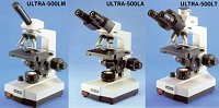
Zenith Ultra-500LT Trinocular Laboratory Microscope
Supplied with Seidentopf Trinocular Head
Full sized laboratory standard instrument of modern ergonomic design,
robust reliable construction and high optical performance. Ideal for use
in schools, colleges and professional laboratories over a wide range of
biological, medical, veterinary, bacteriological and agricultural
applications. Highly recommended.
Optical microscopes
Main article: Optical microscope
Optical microscopes, through their use of visible wavelengths of light, are the simplest and hence most widely used type of biology and geology.
Optical microscopes use refractive lenses, typically of glass and occasionally of plastic, to focus light into the eye or another light detector. Typical magnification of a light microscope is up to 1500x with a theoretical resolution of around 0.2 micrometres or 200 nanometers. Specialised techniques (e.g., scanning confocal microscopy) may exceed this magnification but the resolution is an insurmountable diffraction limit.
Various wavelengths of light are sometimes used for special purposes, for example, in the study of biological tissue.[2] Ultraviolet light is used to illuminate the object being viewed in order to excite a fluorescent dye which then emits visible light. Infrared light is used to study thick slices of biological tissue because infrared light's low diffraction coefficient permits viewing deeper into tissue.
Other microscopes which use electromagnetic wavelengths not visible to the human eye are often called optical microscopes. The most common of these, due to its high resolution yet no requirement for a vacuum like electron microscopes, is the x-ray microscope.
- Magnification x40-x1000, (x1600) achievable by using optional x16
DIN flatfield eyepieces
- Paired x10 DIN standard high eyepoint widefield eyepieces, field
18mm
- DIN standard Parfocal, parcentred achromatic objectives x4
(0.10),x10 (0.25),40R (0.65),x100R (1.25) oil immersion
(R=retractable)
- Bright field ABBE condenser (N.A. 1.25) with iris diaphragm and
filter carrier on fully focussing Rack and Pinion Substage
- Build in 230v, 6v 20w halogen illumination with continuously
variable rotary brightness control
- Co-axial coarse and fine focussing with indexed scale and
adjustable focus tension
- Adjustable focus-stop to prevent damage to glass microslides and
objective lenses
- Smooth action x-y mechanical stage 125x130mm with co-axial drop
controls and an adjustable spring arm to accommodate slides of
different sizes
- Viewing Head inclined 30degrees Rotatable 360degrees with full
inter-pupillary adjustment Magnification factor x1
- Quadruple objective turret on sealed ball bearing race
- Complete with dust cover
- Supplied in polystyrene pack
- Dimensions 215x170x360mm
- Weight 6.5kg
| Accessories |
| WF-5 |
x5
DIN Eyepiece |
| H-7 |
x7
DIN Huyghenian Eyepiece |
| WF-16 |
x16
DIN Widefield Eyepiece |
| ME-10 |
x10
DIN Widefield Measuring Eyepiece |
| OMC-27 |
x20
DIN Achromatic Objective |
| PL-4 |
x4
DIN Planachromatic Objective |
| PL-10 |
x10
DIN Planachromatic Objective |
| PL-25 |
x25
DIN Planachromatic Objective |
| PL-40 |
x40R
DIN Planachromatic Objective |
| PL-100 |
x100R(Oil)
DIN Semi-Plan. Objective |
| TH-2 |
Trinocular
Attachment |
| CA-5 |
SLR
Camera Adaptor (for use with TH-2) |
| BS-2 |
Plano
concave mirror in mount |
| SB-25 |
Replacement
6v 20w Halogen Bulb |
|
Product Code: UTLRA-500LT
build_table('etistore',array('heultra500lt')); ?>
Microscope
From Wikipedia, the free encyclopedia
Jump to: navigation, search
A microscope (Greek: μικρόν (micron) = small + σκοπεῖν (skopein) = to look at) is an instrument for viewing objects that are too small to be seen by the naked or unaided eye. The science of investigating small objects using such an instrument is called microscopy. The term microscopic means minute or very small, not visible with the eye unless aided by a microscope. The microscopes used in schools and homes trace their history back almost 400 years.
The first useful microscope was developed in the Netherlands in the early 1600s.[1] Three different eyeglass makers have been given credit for the invention: Hans Lippershey (who also developed the first real telescope); Hans Janssen; and his son, Zacharias. The coining of the name "microscope" has been credited to Giovanni Faber, who gave that name to Galileo Galilei's compound microscope in 1625. (Galileo had called it the "occhiolino" or "little eye".)
The most common type of microscope—and the first to be invented—is the optical microscope. This is an optical instrument containing one or more lenses that produce an enlarged image of an object placed in the focal plane of the lens(es). There are, however, many other microscope designs.
Contents [hide]
1 Types
1.1 Optical microscopes
1.2 Electron microscopes
1.3 Scanning probe microscope
1.4 Other microscopes
2 External links
An optical light microscope
A stereo microscope is often used for lower-power magnification on large subjects."Microscopes" can largely be separated into three classes, optical theory microscopes, electron microscopes and scanning probe microscopes.
Optical theory microscopes are microscopes which function through the optical theory of lenses in order to magnify the image generated by the passage of a wave through the sample. The waves used are either electromagnetic in optical microscopes or electron beams in electron microscopes. The types are the Compound Light, Stereo, and the electron microscope.
Optical microscopes
Main article: Optical microscope
Optical microscopes, through their use of visible wavelengths of light, are the simplest and hence most widely used type of biology and geology.
Optical microscopes use refractive lenses, typically of glass and occasionally of plastic, to focus light into the eye or another light detector. Typical magnification of a light microscope is up to 1500x with a theoretical resolution of around 0.2 micrometres or 200 nanometers. Specialised techniques (e.g., scanning confocal microscopy) may exceed this magnification but the resolution is an insurmountable diffraction limit.
Various wavelengths of light are sometimes used for special purposes, for example, in the study of biological tissue.[2] Ultraviolet light is used to illuminate the object being viewed in order to excite a fluorescent dye which then emits visible light. Infrared light is used to study thick slices of biological tissue because infrared light's low diffraction coefficient permits viewing deeper into tissue.
Other microscopes which use electromagnetic wavelengths not visible to the human eye are often called optical microscopes. The most common of these, due to its high resolution yet no requirement for a vacuum like electron microscopes, is the x-ray microscope.
Electron microscopes
Main article: Electron Microscope
MicroscopesAcronyms in microscopy
Angular resolution
Bright field microscopy
Condensed Matter Physics
Confocal microscopy
Dark field microscopy
Electron Microscope
Fluorescence interference contrast microscopy
Fluorescence microscope
Microscope image processing
Microscopy
Optical Microscope
 Zenith
Microscopes
Zenith
Microscopes